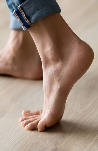
Avascular Necrosis/Osteonecrosis of the Knee
Avascular necrosis of the knee also known as osteonecrosis occurs when blood supply to the thigh bone, also known as the femur or the shin bone, known as the tibia, is disrupted. Bone cells are living, functioning tissue and require a blood supply just like any other tissue in our body. When it does not receive this consistent blood supply, bone begins to degenerate, causing a breakdown in the joint including very painful arthritis.
Osteonecrosis most often occurs in the medial femoral condyle – a part of the thigh bone that meets the knee on the inside of the joint that acts as a primary weight bearing structure in the knee. It can also occur on the outside of the thigh bone or even the top of the shin bone. No matter where it occurs, it can cause significant pain and disability. An early diagnosis does increase treatment options to slow progression, however, as the disease progresses, surgery is almost always necessary to restore function in the joint.
The Causes of Osteonecrosis in the Knee
Anything that affects blood flow to the bone tissue of the knee can cause osteonecrosis. In many cases, it may not be entire clear what caused the blood supply to be interrupted. However, we know of certain risk factors including advanced age, injury to the knee, long-term oral corticosteroid use, obesity, kidney transplantation, alcohol use and more. Regardless of the cause, the progression of treatment will be similar. However, if the cause is a modifiable lifestyle factor, any successful treatment must include a significant change in habits.
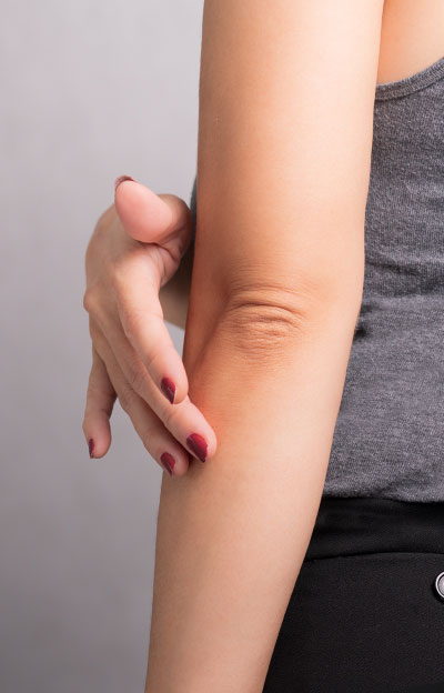
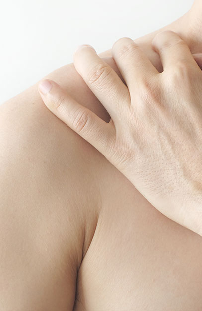
Signs and Symptoms of Avascular Necrosis/Osteonecrosis
Osteonecrosis is a progressive condition and patients will typically feel pain on the inside of the knee as one of the first symptoms. There is no telling when this will occur and can happen gradually or suddenly, especially after an injury occurs. As the disease gets worse, normal activity, even standing, becomes more difficult. Most patients will experience swelling in the knee, sensitivity around the joint and poor range of motion. This can occur over the course of a few months to a year or even more. When caught early, osteonecrosis of the knee can be treated more effectively.
There are four stages of osteonecrosis, with stage four being a complete collapse of the joint space, causing exceptional pain and disability.
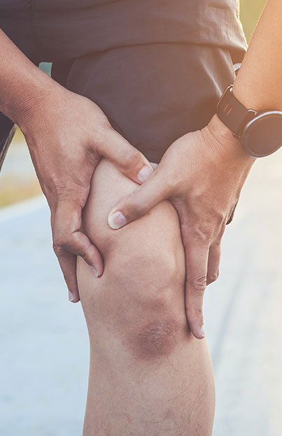
Diagnosing Avascular Necrosis/Osteonecrosis
The diagnosis for avascular necrosis starts with a physical examination to check range of motion, stability, pain, injured tendons or ligaments and more. Imaging is necessary to confirm the diagnosis and includes x-rays and in some cases MRI. MRIs are usually more sensitive to earlier stage cases of the condition.
Treatment for Avascular Necrosis/Osteonecrosis
Treatment will largely depend on the length of progression and the severity of the degradation of the knee. If caught early enough, medication, certain exercises, physical therapy, and the reduction of weight on the knee through modified activities and even crutches can all help slow the progression of the disease and even allow the bone to begin healing.
In particularly severe or advanced cases, or where nonsurgical treatment has failed to offer significant improvement, surgical treatment may be necessary. There are several treatments available to most patients.
- Core Decompression: If the osteonecrosis is diagnosed relatively early and the patient is referred to surgery, core decompression may be very successful. This is where one large and/or a few small holes are drilled into the bone to relieve pressure and stimulate the growth of new blood vessels in the bone area.
- Arthroscopic Debridement and Micro-fracture: Another surgical option includes arthroscopic debridement and micro fracture in which the damaged bone and cartilage is removed from the joint. This is combined with drilling small holes in the bone to improve blood flow to the area.
- At this point, new bone and cartilage may need to be grafted onto the existing healthy bone structure. This is done to build up the damaged area of the joint with healthy bone and cartilage from a donor or the patient’s own body. Synthetic bone grafts may also be considered in some cases
- Autologous Chondrocyte Implantation or ACI involves harvesting cartilage cells, known as chondrocytes, from the patient’s knee. These cells are sent to a laboratory where they are replicated and ultimately implanted back into the knee, where new cartilage is needed. These lab-grown cells go to work by replacing damaged cartilage with healthy cartilage.
- Osteotomy is sometimes employed, where part of the shin bone or thigh bone is removed to mechanically shift weight away from the damaged area of the knee. By shifting this weight, the bone is given additional opportunity to heal. However, this is not an option for many patients.
- In the most severe cases, where the cartilage and bone structure has collapsed and severe arthritis has occurred, a total knee replacement may be necessary. During the knee replacement, Dr. Manning replaces the damaged bone and cartilage with implanted materials that mimic joint function.
Prognosis
The sooner that osteonecrosis is Identified and treated, the more options there are for a successful outcome. However even in the most severe cases, where a total joint replacement is necessary, the outcomes are generally favorable. Most importantly, to minimize the risk of surgery and to maximize outcomes, it is important to choose an experienced orthopedic surgeon such as Dr. Manning to perform surgery.
To learn more and schedule a consultation, we encourage you to contact us.